The role of IgG autoantibodies in CIDP
In patients with CIDP, the binding activity of autoantibodies, including IgG autoantibodies, to the myelin sheath may result in demyelination, subsequent nerve damage, and symptom expression.1,3,4
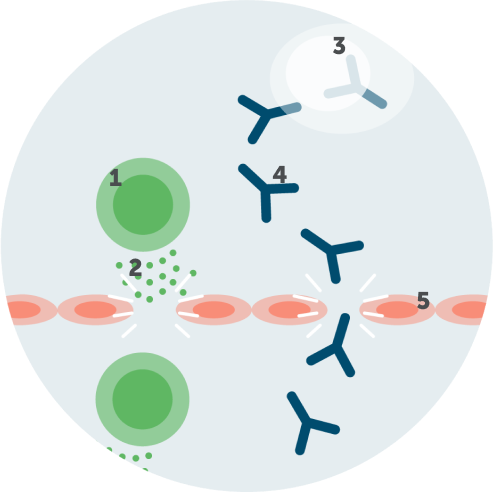
Infiltration of IgG autoantibodies
Activated T cells trigger inflammatory mediators (cytokines and chemokines) to break the blood-nerve barrier, facilitating the passage of autoantibodies, including IgG autoantibodies.1,2,4

1. Activated T cell
2. Cytokines/chemokines
3. Plasma cell
4. IgG autoantibodies
5. Blood-nerve barrier
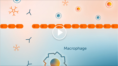
Demyelination due to IgG autoantibodies
IgG autoantibodies bind to the myelin sheath, where they either recruit macrophages directly or activate the complement pathway, which may result in myelin damage.1-4
1. IgG autoantibodies
2. Macrophage
3. Fc receptor
4. Myelin sheath
5. Complement proteins
CIDP=chronic inflammatory demyelinating polyneuropathy; Fc=fragment, crystallized; IgG=immunoglobulin G.
CIDP=chronic inflammatory demyelinating polyneuropathy.
References: 1. Mathey EK et al. J Neurol Neurosurg Psychiatry. 2015;86(9):973-985. doi:10.1136/jnnp-2014-309697 2. Querol LA et al. Neurotherapeutics. 2022;19(3):864-873. doi:10.1007/s13311-022-01221-y 3. Dziadkowiak E et al. Int J Mol Sci. 2022;23:2-13. doi:10.3390/ijms23010179 4. Koike H et al. Neurol Ther. 2020;9:213-227. doi:10.1007/s40120-020-00190-8
